Preface | Introduction to Problem Solving | Problem Sets | Acknowledgments
| E.2. Kinesin <<Previous Problem Next Problem>> | PPDF |
(28 pts) While you're enjoying the “intense” Spring sunshine in Vermont, I thought you might like thinking about some interesting data generated by the use of polarized laser energy, another very intense form of illumination. I've modified the problem from one that appears in The Problem Book (which accompanies Molecular Biology of the Cell by Alberts, et al).
The movement of single motor proteins can be monitored using polarized laser light. Such illumination creates a circular interference pattern with a circular force field that ranges from zero at the center to a few piconewtons at the periphery (about 200 nm from the center). Individual molecules entering this interference pattern are pushed towards the center and "captured" there, unless they possess sufficient kinetic energy to escape. These interference patterns are often called "optical tweezers" because scientists can use them to move molecules about, simply by repositioning the interference patterns. And the actual work accomplished by motility mechanisms can be estimated from the force necessary to escape such constraints. Nifty!!
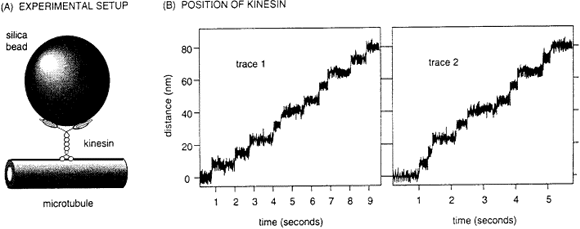
One experiment used optical tweezers to position a kinesin molecule on a microtuble attached to a coverslip (as illustrated below in Fig. A.). The behavior of the kinesin could not be resolved microscopically but its movement was readily tracked by attaching a much larger silica bead to it. When incubated in an appropriate physiological solution, the bead vibrated with the kinesin molecule, due to thermal kinetics, but it did not move away from the center of the interference pattern. When ATP was added to this test set-up, the bead began to migrate away from the center and towards the one end of the microtubule.
As it moved along the microtubule, kinesin encountered the force of the interference pattern, which is thought to simulate the kind of loads born by kinesin in moving vesicles around the cell. Traces of two kinesin molecules along a microtubule are illustrated in Fig. B. Consider these data and answer all the following questions.
|
|
1. (4 pts) What supplies the free energy needed for unidirectional (non-Brownian)
movement along the microtubule?
2. (6 pts) What is the average rate of kinesin movement and what is the
average step length?
3. (8 pts) In each protofilament the tubulin subunits repeat at 8 nm intervals.
Given the average step length and this repeat interval, postulate a detailed
mechanism for the movement of kinesin.
4. ( 4 pts) How many ATP molecules are likely hydrolyzed per step? How
did you arrive at your answer?
5. (8 pts) Towards which end of the microtubule does kinesin move, and
what biochemical constraints might polarize this movement? (Recall that
dynein moves along MT in the opposite direction.)