Studying
Cell Biology
Preface |
Introduction to Problem Solving | Problem
Sets | Acknowledgments
|
E.5.a. Axon Movement <<Previous Problem |
Print
PDF
|
| Next Part> |
| E.5.a. |
| E.5.b. |
|
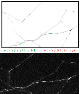
Neurons are elongate, highly polarized cells consisting of a cell body (which contains the nucleus, ER and many organelles), branching projections of the cell body called dendrites, and a very long extension called an axon. Typically, specialized regions called synapses are found at the extremities of the dendrites and axon, and patch-clamp measurements of membrane potentials indicate action potentials are usually propagated along the neuron from the dendritic synapses to the axonal synapse. In addition, the axon exhibits rapid transport of vesicles along its entire length. To investigate the transport of vesicles in the axon, Kaether, C. et al. (2000, Mol. Biol. Cell. 11:1213-1224. PMID10749925) grew hippocampal neurons in culture and examined axonal outgrowths by phase contrast and fluorescence microscopy. Their fluorescent images are displayed in the figures and video below. To follow the movement of axonal vesicles, the gene for a ubiquitous IMP (amyloid precursor protein, APP) was tagged with the gene for yellow fluorescent protein (YFP), and the chimeric gene was expressed in the cultured neurons. The movement of vesicles containing APP-YFP was followed by fluorescence microscopy, and the upper figure below, which is a “negative” image of the one beneath it, highlights two types of tubular vesicle: those moving from left to right (outlined in red) towards the axon tip and those moving from right to left (outlined in green) towards the cell body. After you have become oriented to the figure, click on the image below to start the movie. The video sequence contains 50 frames which were captured at 0.9 sec intervals, for a total filming time of 45 sec. The duration of the QuickTime movie is 5 sec, and hence the vesicular movements are speeded up 9 times. The width of the video frame is about 100 mm, and for reference purposes, the lower axon running diagonally across the frame, with two “red” and two “green” vesicles depicted, is also about 100 mm in length. Play the movie several times, at normal speed and one frame at a time, to follow the movements of the two kinds of vesicles and answer the following questions. [For more information about fast axonal transport, please consult the research article by Kaether, C. et al., 2000, Mol. Biol. Cell. 11:1213-1224. PMID10749925.
|
|||
|
Questions
B. What cytoskeletal organelle likely guides the movement of these
vesicles? C. How could you test this hypothesis? D. How fast are the vesicles moving towards the axon extremities? Towards the cell body? E. What molecular motors might be responsible for the vesicular
movements? Discuss briefly the basis F. How could you test your hypothesis? To examine the results of one experiment designed to identify a molecular motor, turn the page. |
||